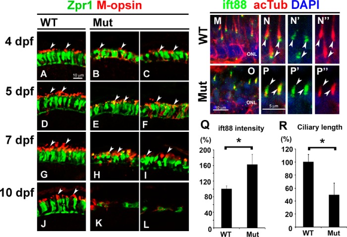
FIGURE 5.
Defect of the opsin transport machinery in the ift122 mutant retina. A–L, photoreceptor cells at 4 dpf (A–C), 5 dpf (D–F), 7 dpf (G–I), and 10 dpf (J–L) in wild-type (A, D, G, and J) and ift122 mutant (B, C, E, F, H, I, K, and L) larvae were immunostained with anti-Zpr1 (green) and anti-M-opsin (red) antibodies. In the 4 dpf wild type, the M-opsin was localized at the outer segments (A). In contrast, the M-opsin signal was widely distributed in photoreceptor cell bodies in ift122 mutant larvae at 4 dpf (B and C). At 5 dpf, ectopic accumulation of M-opsin signal in photoreceptor cell bodies was still observed (E and F), but enrichment of the M-opsin in the outer segment was also observed in mutant photoreceptors. At 7 dpf, the cell bodies of mutant photoreceptors were smaller compared with those of wild-type photoreceptors (H and I). At 10 dpf, only degenerated photoreceptors were observed in most of the photoreceptor layer in the mutant retina (K and L). Arrows indicate outer segments. M–P″, retinal sections were immunostained with an anti-Ift88 antibody (green) in the wild-type (M–N″) and mutant larvae (O–P″) at 4 dpf. Photoreceptor cilia are immunostained with an anti-acetylated α-tubulin antibody (red). Accumulations of the ift88 signals are observed in the photoreceptor cilia of mutant larvae. Q and R, quantification of length and ift88 signal intensity in photoreceptor cilia of ift122 mutant larvae at 4 dpf. Accumulation of ift88 at ciliary tips was significantly increased in ift122 photoreceptor cilia (Q). Photoreceptor cilia stained with acetylated α-tubulin were significantly shorter in ift122 mutant retina (R). Nuclei were stained with DAPI (blue). ONL, outer nuclear layer.

