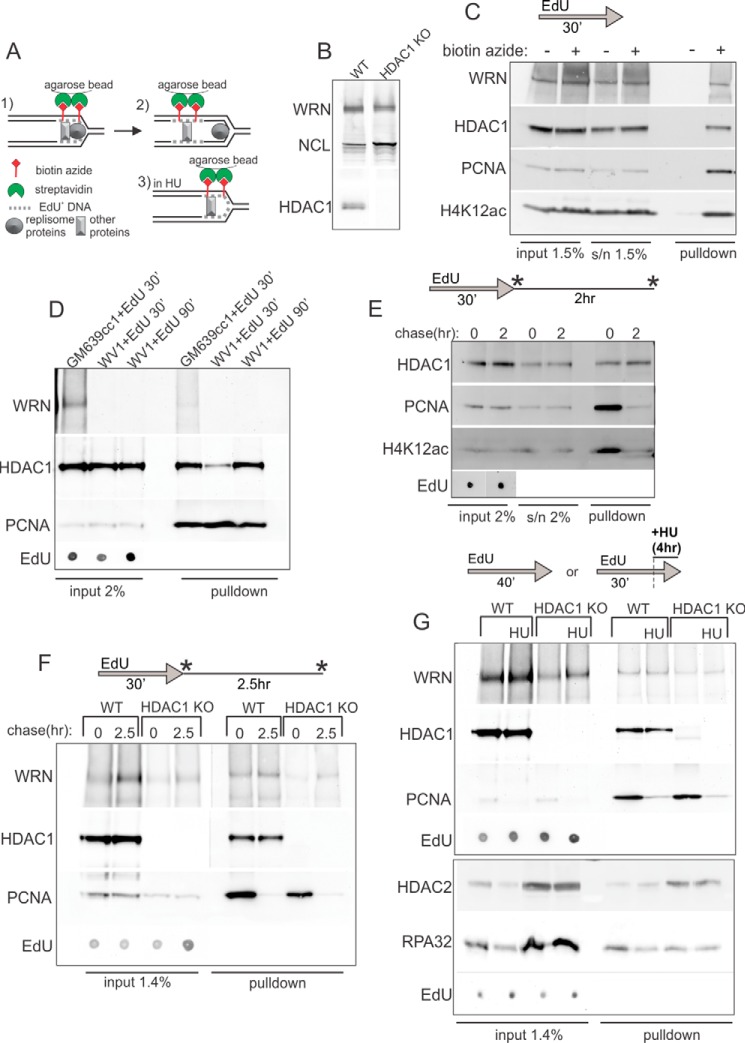
FIGURE 4.
HDAC1 and WRN bind nascent DNA. A, a schematic of iPOND. B, Western blot verification of loss of expression of HDAC1 in the HDAC1 knock-out clone (#17) of RD cells. Nucleolin (NCL) and WRN expression are shown for reference. C, a Western blot of niPOND precipitates from GM639cc1 cells labeled by EdU for 30 min, harvested, and subjected to a Click-iT reaction with and without biotin-azide before lysis and incubation with streptavidin beads. D, Western blots of niPOND performed with GM639cc1 and WV1, WRN-deficient fibroblasts. WV1 cells were labeled with EdU for 30 or 90 min. For EdU, serial 2- or 3-fold dilutions from 0.001% to 0.015% of lysates were loaded on a dot blot and visualized with HRP-conjugated anti-biotin antibody. EdU panels show a representative dilution within a linear range of EdU signal. E, a Western blot of niPOND performed with GM639cc1 cells labeled with EdU for 30 min and harvested immediately or chased for 2 h before harvest. F, a Western blot of niPOND performed with RD cells with intact (WT) or knocked-out (HDAC1 KO) HDAC1 gene. Cells were harvested immediately after an EdU pulse or after a 2.5-h chase. G, a Western blot of niPONDs performed with RD cells labeled with EdU for 30 min or for 15 min followed by a 6-h arrest with 2 mm HU in the presence of EdU. Panels framed in separate boxes come from independent experiments as simultaneous detection of HDAC1 and HDAC2 on one Western blot is not optimal due to their close molecular weights. More RD HDAC1 KO than WT cells were used in panels F and G in order to compensate for their lower percentage of replicating cells. Residual bands larger and smaller than expected for HDAC1 seen in the HDAC1 KO, no-HU lane may be nonspecific or represent cross-reactivity with HDAC2. Asterisks in E and F indicate time points of sample harvest.

