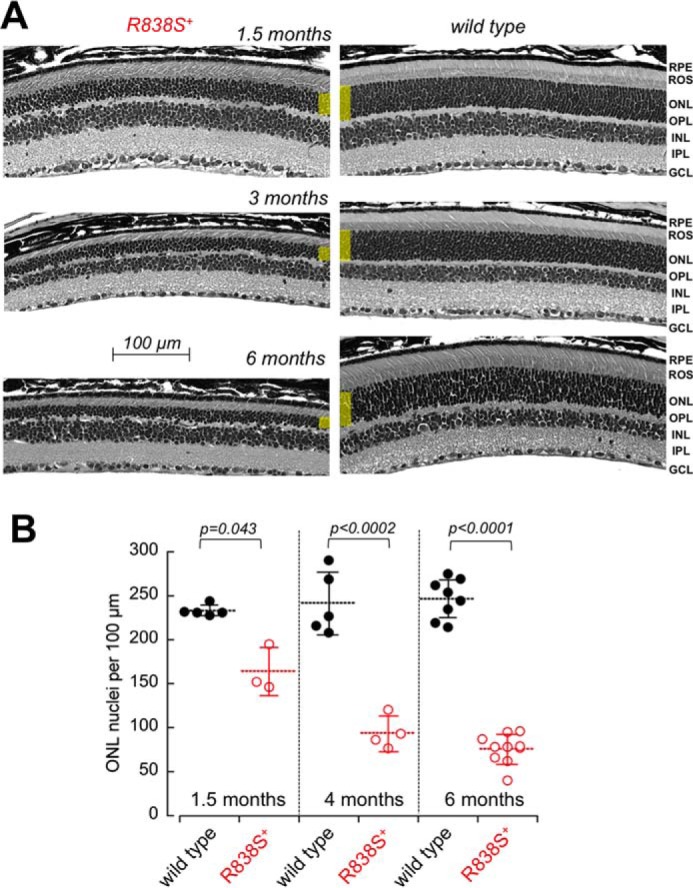
FIGURE 6.

Degeneration of the photoreceptor layer in R838S+ mice. A, the cross-sections of the retinas from the slower line 379 R838S+ at 1, 3, and 6 months of age (left), shown next to their non-transgenic littermates (right). Note the deterioration of the rod morphology and the reduction in their outer nuclear layer thickness (highlighted yellow); RPE, retinal pigment epithelium; OPL, outer plexiform layer; INL, inner nuclear layer; IPL, inner plexiform layer; GCL, ganglion cell layer. B, the progressive loss of average photoreceptor nuclei count per 100-μm retina length in the R838S RetGC1-positive mice (open symbols) and their wild type littermates (closed symbols); dashed horizontal bars, mean average for each group; error bars, standard deviation.
