FIGURE 7.

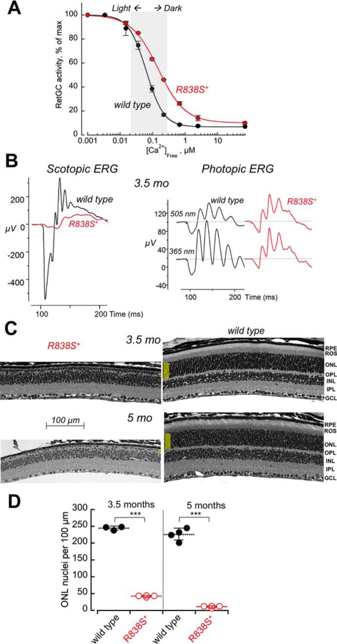
A stronger shift in Ca2+ sensitivity of RetGC1 exacerbates the degenerative phenotype in R838S RetGC1-positive mice. A, calcium sensitivity of cGMP synthesis measured in the retinas from R838S RetGC1 positive (red symbols) and negative (black symbols) littermates of the fast line 362, aged 3 weeks. The assays were conducted as described in the legend to Fig. 2B; the K½Ca from the Hill fit in two independently tested transgenic mice was 148 ± 2 nm compared with the 63 ± 10 nm in four wild type siblings (p < 0.001). B, the waveforms of scotopic (left panel) and photopic (right panel) ERG in 3.5-month-old transgenic (red traces) and wild type (black traces) littermates. C, retinal morphology in line 362 at 3.5 and 5 months of age. Note the extent of reduction of the outer nuclear layer compared with the wild type littermates. D, the loss of photoreceptors in the R838S RetGC1-positive mice (open symbols) and their wild type littermates (closed symbols); horizontal bars, mean average for each group. The average photoreceptor nuclei count per 100 μm length in the transgene-positive retinas was 42 ± 2 compared with the 244 ± 6 in wild type after 3.5 months, and 11 ± 3 versus 226 ± 18 after 5 months (p ≤ 0.0001).
