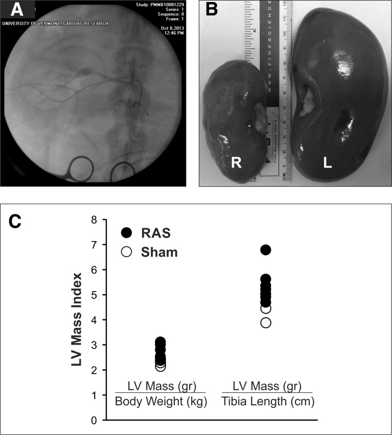
Fig. 2.
Unilateral RAS and LV hypertrophy. A: radiographic contrast image of the right renal artery with a high-grade RAS and poststenosis dilation. B: reduction of right (R) kidney size after 11 wk of RAS. L, left. C: LV mass measured at the end of the study indexed to body weight and tibia length.

