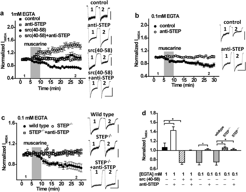
Figure 4. The direction of change in NMDAR function as a consequence of mAchR stimulation is influenced by the intracellular concentration of EGTA.
(a) With 1 mM intracellular EGTA, muscarine application (10 μM; timing indicated by the shaded region) fails to potentiate NMDAR currents (n = 5, 1.07 ± 0.10), but can do so in the presence of anti-STEP (n = 6, 1.43 ± 0.09, P < 0.05 compared with muscarine). Conversely, with 1 mM EGTA, muscarine depresses NMDAR currents in the presence of Src (40–58) (n = 5, 0.74 ± 0.04, P < 0.05 compared with muscarine) but not in the presence of both Src(40–58) and anti-STEP (n = 6, 1.00 ± 0.01). (b) With 0.1 mM intracellular EGTA, the depression of NMDAR currents by muscarine (n = 5, 0.74 ± 0.05) is inhibited by anti-STEP (n = 7, 1.02 ± 0.02). (c) With 0.1 mM intracellular EGTA, muscarine application depresses NMDAR currents in neurons from wild type mice (n = 7, 0.75 ± 0.07) but not in neurons from STEP KO mice (n = 7, 1.05 ± 0.02, p > 0.05 compared with baseline). Anti-STEP had no effects on NMDAR currents in STEP KO mice (n = 5, 1.02 ± 0.02, P > 0.05 compared with STEP KO mice without anti-STEP). (d) Summary bar graph of data in (a–c). *Indicates p < 0.05. Values represent the average of peak current amplitudes recorded between 20–25 min normalized to the average of peak current amplitudes recorded during the first 5 min of recording, prior to muscarine application. Calibration bars: 3s; (a) control 100 pA, anti-STEP 300 pA, Src(40–58) 200 pA, Src(40–58) + anti-STEP 200 pA. (b) control 300 pA, anti-STEP 400 pA. (c) wild type 600 pA, STEP−/− 200 pA, STEP−/− + anti-STEP 200 pA.

