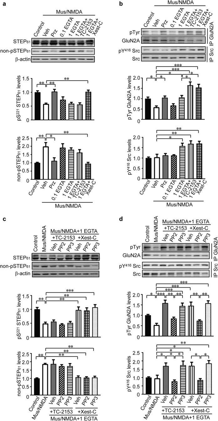
Figure 6. Tyrosine phosphorylation of GluN2A is regulated by STEP61 and Src activities.
(a) Primary hippocampal neurons were pretreated with various inhibitors (veh: 0.1% DMSO; pirenzepine: 10 μM; EGTA/AM: 0.1 or 1 mM; TC-2153: 1 μM; xestospongin C: 10 μM) for 30 min, followed by co-stimulation of muscarine (10 μM) and NMDA (50 μM) for 10 min. Neuronal lysates were probed with anti-STEP (23E5) and anti-non-phospho-STEP61 antibodies. Phospho- and non-phospho- STEP61 levels were normalized to total STEP61 protein levels and then to β-actin as loading control. (b) GluN2A and Src were immunoprecipitated from treated lysates using specific antibodies, followed by probing with anti-Tyr, anti-pY416 Src and pan-protein antibodies, respectively. Phospho-protein levels were normalized to pan-protein levels. (c) In addition to the inhibitors used in (a), PP2 (10 μM) and PP3 (10 μM) were added to ECS for stimulation. Phospho- and non-phospho- STEP61 levels were assessed. (d) After treatments, phosphorylation of GluN2A and Src were measured on immunoprecipitated samples. Phospho-protein levels were normalized to pan-protein levels. All data were expressed as mean ± SEM. Statistical significance was determined using one-way ANOVA with post hoc Tukey test (*P < 0.05, **P < 0.01, ***P < 0.001, n = 5).

