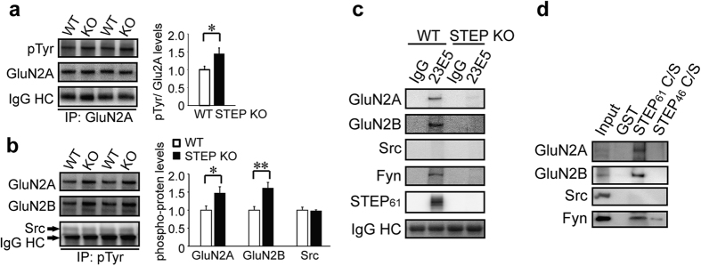
Figure 7. STEP binds to and dephosphorylates GluN2A.
Synaptosomal fractions from WT and STEP KO mouse hippocampus were used for immunoprecipitation with anti-GluN2A (a) or anti-phophos-Tyr (b), followed by immunoblotting with anti-phopho-Tyr or anti-GluN2A, anti-GluN2B and anti-Src, respectively. Phospho-protein levels were normalized to total proteins, repectively. All data were expressed as mean ± SEM. Statistical significance was determined using two-tailed Student’s t test (*P < 0.05, **P < 0.01, n = 6). (c) STEP was immunoprecipitated from WT and STEP KO hippocampal lysates using anti-STEP (23E5) antibody. Potential interacting proteins were verified using selective antibodies indicated in the figure. Representative blots were shown from three independent replicates. (d) GST-STEP fusion proteins were bound to glutathione-sepharose 4B beads and incubated with hippocampal lysates. Co-purified proteins were verified using selective antibodies indicated in the figure. Representative blots were shown from three independent replicates.

