
Figure 1.
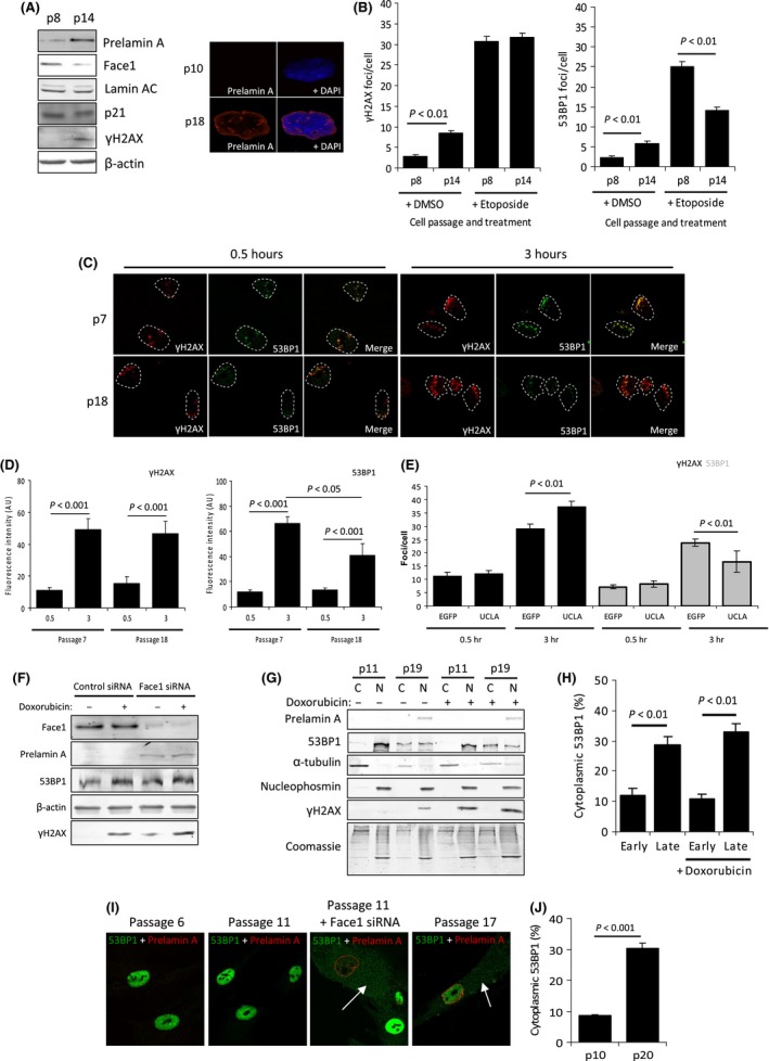
Prelamin A in aged VSMCs prevents 53BP1 recruitment to DNA damage by inducing cytoplasmic accumulation. (A) (Left) WB showing increased prelamin A in aged (p14) VSMCs compared to early (p8) VSMCs that occurs concomitantly with a decrease in Face1. Levels of mature lamin AC and p21 are not markedly different, whereas γH2AX is increased. The data shown are from 35F VSMC isolate, but we also detect prelamin A accumulation and increased γH2AX in two other VSMC isolates (Fig. S1). All experiments were repeated a minimum of 3 times. (Right) IF image of a proliferative early passage (p10) VSMC and a nonproliferative late passage (p18) VSMC stained for prelamin A (red) and DAPI (blue). (B) Enumeration of γH2AX (left) and 53BP1 (right) foci in p8 and p14 VSMCs treated with DMSO or etoposide. n > 200 cells per treatment taken from 3 independent experiments. Standard errors are shown. (C) IF showing γH2AX (red) and 53BP1 (green) in p7 and p18 VSMCs treated with microirradiation and left to recover for 0.5 or 3 h. About 84% of p18 VSMCs were positive for prelamin A, and no prelamin A was detected in p7 VSMCs (data not shown). (D) Quantification of C. n > 100 cells per treatment taken from 3 independent experiments. (E) IF analysis of γH2AX and 53BP1 foci in p8 control (EGFP) or expressing prelamin A (UCLA) VSMCs treated with etoposide for 3 h. n = 200 cells per treatment taken from 3 independent experiments. (F) Whole cell lysate WB taken from early passage VSMCs treated with control or Face1 siRNA and −/+ doxorubicin treatment. Levels of prelamin A were increased, but total 53BP1 protein level did not change. (G) WB of cell fractionation analysis showing cytoplasmic 53BP1 accumulates in p19 VSMCs and that this coincides with prelamin A expression (not seen in p11 VSMCs). α‐tubulin and nucleophosmin are shown as controls for cytoplasmic (C) and nuclear (N) fractions, respectively. Doxorubicin did not affect this accumulation. (H) Quantification of G. Data were taken from a minimum of 3 separate experiments. (I) IF of 53BP1 (green) cytoplasmic accumulation in VSMCs that have accumulated prelamin A (red). p6 and p11 VSMCs had undetectable levels of prelamin A, but induced expression of Prelamin A with Face1 siRNA (third panel) caused an increase in levels of cytoplasmic 53BP1 and reduced nuclear levels. This change is also evident in p17 VSMCs that have naturally accumulated prelamin A. White arrows indicate cytoplasmic 53BP1. Nuclei have been stained with DAPI. (J) Quantification of fluorescence measurements of cytoplasmic and nuclear 53BP1 in p10 and p20 VSMCs. n > 100 cells from 3 separate experiments.