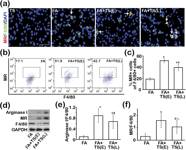
Figure 7. Tanshinone IIA promotes M2 macrophages polarization in the folic acid injured kidney.
(a) Representative micrographs showing immunofluorescent staining of kidney specimens procured on day 7 for F4/80 and mannose receptor (MR). Sections were counterstained with 4′,6-diamidino-2-phenylindole (DAPI). Scale bar = 50 μm. White arrows indicate the dually positive cells in the sections. (b) Flow cytometry analysis of isolated single kidney cells for cells positive for MR and F4/80; cells were gated on F4/80+ macrophages. (c) The percentage of F4/80+MR+ cells among all F4/80+ cells as determined by flow cytometry. *P < 0.001 vs FA; #P < 0.001 vs FA+TS(E); (n = 9). (d) Representative immunoblots of Arginase I, MR, and F4/80 in cortical kidney lysates prepared on day 7. (e) and (f) Relative abundance of Arginase I (e) and MR (f) in immunoblots expressed as densitometric ratios of Arginase I/F4/80 or MR/F4/80. *P < 0.001 vs FA; $P = 0.041 vs FA+TS(E); @P = 0.002 vs FA; △P = 0.029 vs FA+TS(E); (n = 9).

