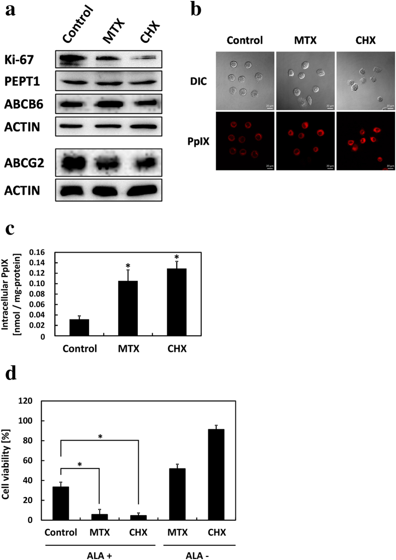
Figure 6. Porphyrin metabolism changed in cancer cells on addition of methotrexate (MTX) or cycloheximide (CHX).
MTX was cultured with 10 μM and CHX was cultured with 10 μg/ml for 48 h. (a) Protein expression was detected by Western blotting. (b) Confocal laser scanning microscopy images of PpIX accumulation. Scale bar, 20 μm. (c) PpIX accumulation was measured by HPLC. n = 3. *p < 0.005, compared with control. (d) Effect of ALA-PDT on cell viability. Cells were incubated with 1 mM ALA for 24 h and then exposed to 1080 mJ/cm2 light for 5 min. Cell viability was determined with trypan blue. Cell viability was normalized by untreated control samples. n = 3. *p < 0.003. All bars represent standard deviation (SD).

