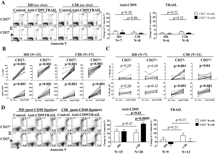
Figure 2. Increased sensitivity to Fas-mediated apoptosis in anti-CD40-activated CD27+ B-cells in cirrhosis.
(A) Purified CD27+ and CD27− B-cells from HD and CIR were incubated with agonistic anti-CD95 mAb (CH11) and recombinant TNF-related apoptosis-inducing ligand (rTRAIL) for 18 hours and then labeled with Annexin-V-FITC and propidium iodide (PI). Cumulative data showing the change in % Annexin-V+ using by subtracting the value for percentage of Annexin-V-positive cells in culture medium alone from the value for percentage of apoptosis in a replicate culture containing agonistic anti-CD95 mAb or rTRAIL. Error bars reflect standard deviation. All comparisons tested with Wilcoxon test. (B) Purified CD27+ and CD27− B-cells from HD and CIR were incubated with agonistic anti-CD40 mAb for 48 hours. The data represent the frequency and geometric mean fluorescence (gMFI) of CD95 on CD27+ and CD27− B-cells ex vivo and after CD40 ligation. All comparisons tested with Wilcoxon test. (C) The frequency of TRAIL-R1 and TRAIL-R2 on CD27+ and CD27- B-cells ex vivo and after CD40 ligation. (D) After CD40 ligation, CD27+ and CD27− B-cells were incubated with agonistic anti-CD95 mAb (CH11) and rTRAIL for an additional 18 hours and then labeled with Annexin-V-FITC and propidium iodide (PI). Cumulative data showing the change in % Annexin-V+ using by subtracting the value for percentage of Annexin-V-positive cells in culture medium alone from the value for percentage of apoptosis in a replicate culture containing agonistic anti-CD95 mAb or rTRAIL. Error bars reflect standard deviation. All comparisons tested with Wilcoxon test.

