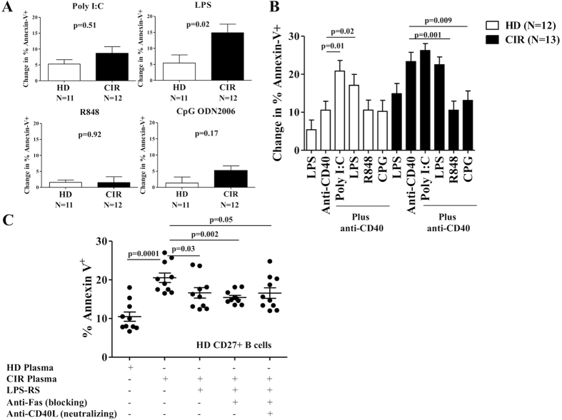
Figure 5. TLR-activation modulates Fas-sensitivity in cirrhotic CD27+ B-cells.
(A) CD27+ B-cells from HD and CIR were incubated with poly(I:C), LPS, R848 and CpG ODN2006 for 48 hours. After TLR ligation, CD27+ B-cells were incubated with agonistic anti-CD95 mAb (CH11) for an additional 18 hours and then labeled with Annexin-V-FITC and PI. Cumulative data showing the change in % Annexin-V+ using by subtracting the value for percentage of Annexin-V-positive cells in culture medium alone from the value for percentage of apoptosis in a replicate culture containing agonistic anti-CD95 mAb. (B) CD27+ B-cells from HD and CIR were incubated with anti-CD40 plus poly(I:C), LPS, R848 and CpG ODN2006 for 48 hours. After CD40/TLR ligation, CD27+ B-cells were incubated with agonistic anti-Fas mAb (CH11) for an additional 18 hours and then labeled with Annexin-V-FITC and PI. Cumulative data showing the change in % Annexin-V+ using by subtracting the value for percentage of Annexin-V-positive cells in culture medium alone from the value for percentage of apoptosis in a replicate culture containing agonistic anti-CD95 mAb. (C) Impact of blocking anti-Fas mAb, blocking anti-CD40L mAb and TLR4 antagonism (inhibitory Lipopolysaccharide Rhodobacter sphaeroides, LPS-RS) on the expression of Annexin-V on healthy donor CD27+ B-cells co-cultured with CIR plasma for 18 hours. Representative data from three separate experiments with different B-cell donors are shown. Error bars reflect standard error of the mean. Statistical comparisons done by Wilcoxon test.

