Fig. 1.

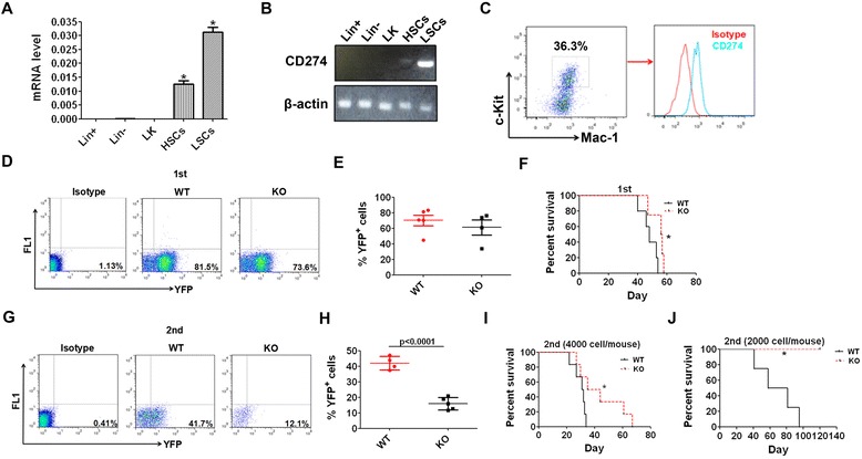
CD274 is highly expressed in LICs and promotes AML development. a, b CD274 expression levels were determined in mouse LICs, HSCs, and other hematopoietic cells (LK, Lin−, and Lin+ cells) by real-time RT-PCR or semi-quantitative PCR. c The expression of CD274 in mouse LICs was examined by flow cytometric analysis. d, e The frequencies of WT and CD274-null leukemia cells (YFP+) in the peripheral blood in primary recipient mice 3 weeks post-transplantation were analyzed. Normal mouse bone marrow cells were used as background (bg) fluorescence control. Representative flow cytometric plots (d) and quantitative results (e) are shown (n = 4–5). f MLL-AF9-induced WT and CD274-null hematopoietic stem/progenitors was transplanted into recipient mice, followed by the analysis of overall survival upon the primary transplantation (n = 4–5). g, h The frequencies of WT and CD274-null leukemia cells in the peripheral blood were determined three weeks after the secondary transplantation. Normal mouse bone marrow cells were used as bg fluorescence control. Representative flow cytometric plots (g) and quantitative results (h) are depicted (n = 4–5). i, j Representative results of the overall survival of the recipient mice receiving 4000 (i) or 2000 (j) WT or CD274-null leukemia cells upon the secondary transplantation (n = 6 for i, and n = 4–5 for j). (*p < 0.05)
