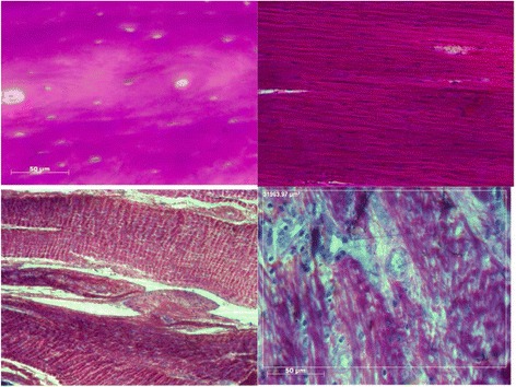
Fig. 8.

Top left: Histology photograph showing DCB before implantation. Top right: remodelled DCB §showing aligned cell infiltration. Bottom left: Well organised collagen fibres within DCB showing crimp. Bottom right: Less organised remodelling in DCB showing vascularisation and collagen fibre formation
