Fig. 2.

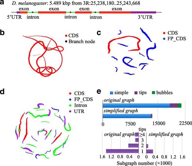
Comparison between the traditional de Bruijn graph and the codon-based de Bruijn graph. a The basic information of FBgn0039298 gene in D. melanogaster used for generating the subfigures (b)–(d). The elements are marked with different colors to highlight the gene structure (UTR: purple, intron: green, exon: red). b Traditional de Bruijn graph based on simulated DNA-seq reads on CDS regions. c Codon-based de Bruijn graph based on simulated RNA-seq reads. Each dot denotes a kmer node. The nodes belonging to CDS are shown in red. The false CDSs translated from the wrong frames of the gene are shown in blue. d Codon-based de Bruijn graph based on simulated DNA-seq reads on the whole gene. The nodes belonging to CDSs are shown in red. The false CDSs translated from the wrong frames of the gene are shown in blue. The false CDSs translated from introns and UTRs are shown in green and purple, respectively. e Comparison between the two codon-based de Bruijn graphs before and after tips trimming and bubble merging using a real RNA-seq dataset (ERR188040). “Simple” indicates subgraphs without tips and bubbles; “tips” indicates subgraphs containing tips; “bubbles” indicates subgraphs containing bubbles and/or tips
