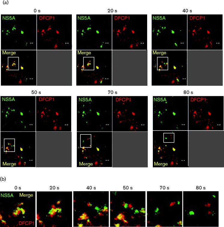
Fig. 7.
Transient association of nascent HCV RNA replication complexes and DFCP1 imaged by live cell microscopy. Huh7 cells were transfected with a mCherry–DFCP1 expression plasmid and at 48 h post-transfection cells were infected with Jc1–GFP (0.5 f.f.u. per cell). At 24 h p.i., live cell imaging was performed and movies of infected cells captured. (a) Representative images captured at the time points indicated. Bars, 2 μm. (b) A montage of the images indicated in the white squares in (a), captured at the times indicated.

