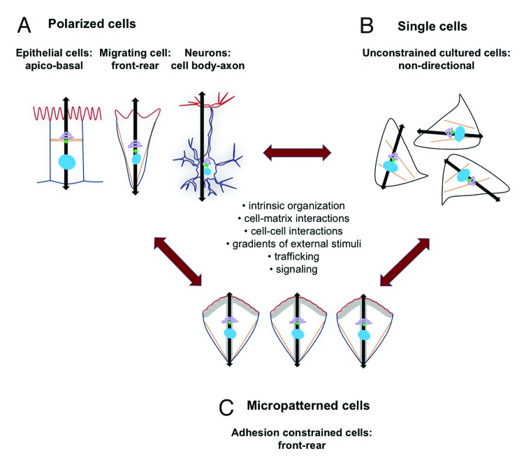
Figure 1. Model of cell polarity and internal cell organization. Cell polarity may be regarded as a gradual change in cell appearance that is regulated by a complex interplay between intrinsic cell organization, cell-matrix interactions, cell-cell interactions, gradients of external stimuli, trafficking pathways and signal transduction cascades. (A) Polarity in cellular organization is observed in epithelial cells (apico-basal polarity), in migrating cells (front-rear polarity) and neurons (cell body- axon polarity). The corresponding polarity axes are represented with black flashes. The nucleus, centrosome and Golgi apparatus are often oriented along this axis. Further, different membrane domains with distinct and mutual exclusive lipid compositions are characteristic for cell polarization (represented schematically with a red and blue line). Nuclei are blue, the Golgi apparatus is purple, the centrosome is green and stress fibers are in orange. (B) Single cells in unconstrained culture condition show an intrinsic cell organization that is however non-directional. Since the internal polarity axes of single cells are not aligned, cells are considered non-polarized. No distinct membrane domains have been observed in unconstrained cultured cells. (C) Micropatterned cells reveal a constant shape due to restrained adhesion that mimic, in vitro, a simplified restriction in space typical for cells in body tissues. On micropatterns, the cell internal polarity axis is aligned along a front-rear orientation.

An official website of the United States government
Here's how you know
Official websites use .gov
A
.gov website belongs to an official
government organization in the United States.
Secure .gov websites use HTTPS
A lock (
) or https:// means you've safely
connected to the .gov website. Share sensitive
information only on official, secure websites.
