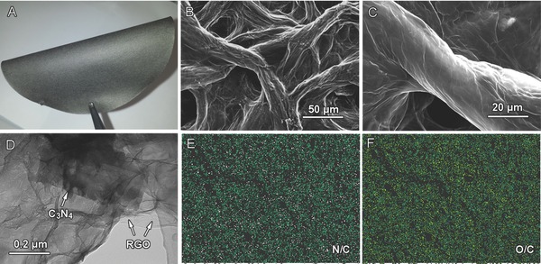
Figure 1.

Typical morphological characterizations of G‐C3N4: A) optical image; B,C) scanning electron microscopy images; D) transission electron microscopy image; E,F) energy dispersive spectrometer element mapping taken from (C) showing distribution of N (pink), O (yellow), and C (green) atoms inside G‐C3N4.
