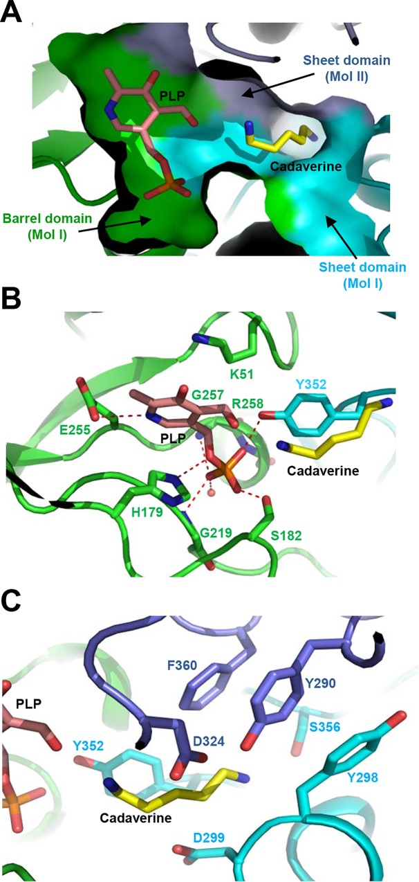
Fig 2. Cofactor and substrate binding mode of SrLDC.
(A) A surface model of the active site of SrLDC. The SrLDC structure complexed with PLP/cadaverine is presented with a surface model with colors of green, cyan, and light-blue for the barrel domain and the sheet domain of one monomer and the sheet domain of the other monomer, respectively. The bound PLP and cadaverine are shown as stick models with salmon and yellow colors, respectively. (B) PLP binding mode of SrLDC. The SrLDC structure complexed with PLP/cadaverine is presented with a cartoon diagram with the same color scheme as in (A). The residues involved in the PLP binding are shown as stick models and labeled. The hydrogen bonds involved in the PLP binding are shown as red-colored dotted lines. The bound PLP and cadaverine are shown as stick models with salmon and yellow colors, respectively. (C) Substrate binding mode of SrLDC. The SrLDC structure complexed with PLP/cadaverine and residues involved in the substrate binding are presented as (B).

