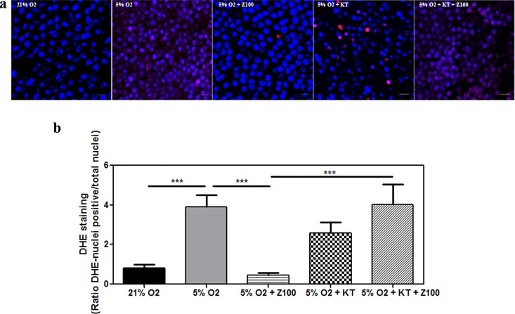
Fig 5. PDE inhibition ameliorated the effects of hypoxia on superoxide release in cultured porcine retinas.
Retinal explants were incubated under normoxia (21% of oxygen) or mild hypoxia conditions (5% of oxygen) for 24h with dimethyl sulfoxide (DMSO), Zaprinast and KT5823 alone or combined. Superoxide radical generation was assessed with the oxidative fluorescent dye dihydroethidium (DHE) in the outer nuclear layer (photoreceptors) as described in Material and Methods. Confocal laser scanning micrographs showing DHE imaging of superoxide accumulation (red). Scale bar: 10 μm (a) in Hoescht 33342-counterstained of live retinal explants, Bar graphs showing the quantification of superoxide (b). Values are the mean ± SEM of five different cultures. Values that are significantly different are indicated by asterisks ***P < 0.001 (linear regression models of mixed effects). Z100: 100 nM Zaprinast; KT: 1 μM KT5823; TAC, total antioxidant capacity.

