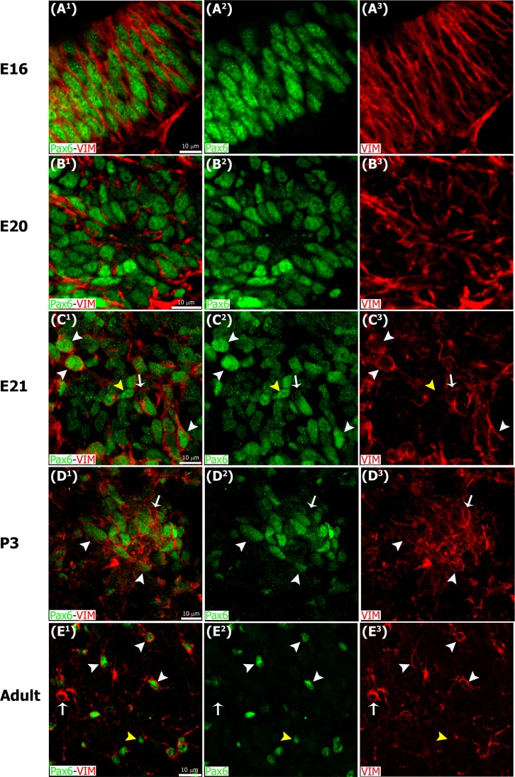
Fig 3. Pax6/Vimentin double immunostaining reveals dynamic changes in the organization of precursor cells throughout pineal ontogeny.
Panels include higher magnification images of the insets shown at 60x and 40x in Figs 1 and 2 immunostained for Pax6 (green) and vimentin (VIM, red). (A1-A3) In the earliest stages Pax6/VIM double-positive precursor cells display a radial distribution. (B1-D3) After fusion of the neuroepithelium, Pax6/VIM+ cells are arranged mainly as rosette-like structures. (E1-E3) In the adult pineal gland (PG) individual cells positive for Pax6 and/or vimentin are dispersed throughout the parenchyma. White arrowheads: Pax6high/VIMhigh cells. Yellow arrowheads: Pax6high/VIMlow cells. White arrows: Pax6low/VIMhigh cells. (A1-E3) 3x, 4x, 3x, 2.5x and 2.6x digital zooms of the insets shown in Figs 1B2–1B4, 1F2–1F4 and 1G2–1G4 and 2A2–2A4 and 2C2–2C4, respectively; scale bar: 10 μm. E, embryonic day. P, postnatal day.

