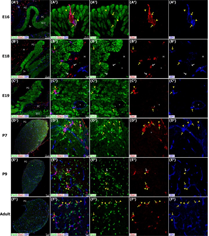
Fig 5. Microglia colonize the pineal gland neuroepithelium very early in development and persist across the lifespan, showing affiliation with Pax6+ cells.
Microglia in the developing and adult pineal gland (PG) exhibit an ‘activated’ morphology. Panels show images of confocal microscopy of PG sections immunolabeled for Pax6 (green) and the microglial marker Iba1 (red), and stained with the isolectin IB4 (IB4, blue) to reveal blood vessels (v) and a subpopulation of microglial cells. Iba1+ microglia were present in the meninges (M) and choroid plexus, and appeared to enter the PG from both sources early in development. In a few cases microglial cells were seen phagocytosing Pax6+ precursor cells (yellow arrowheads) in early postnatal development (A1-A5), but this was most common in the postnatal and adult PG. Yellow arrows show Iba1/IB4 double-positive cells. The tight spatial relationship between macrophages and blood vessels suggests that microglia might also colonize the PG through the vasculature (asterisk). White arrows: microglial cells positive for Iba1 and negative for IB4 (red). (A1, B1) 20x; (C1, E1) 1.6x and 1.2x digital zooms from 10x images, respectively; (D1 and F1) 10x; scale bar: 75 μm. (A2-A5, B2-B5, C2-C5, D2-D5, E2-E5) 3x, 1.4x, 1.7x, 1.5x and 1.4x digital zooms from 60x images, respectively; (F2-F5) 1.5x enlargements from a 40x image; scale bar: 10μm. E, embryonic day. M, meninges. P, postnatal day. PC, posterior commissure. SCO, subcommissural organ. 3v, third ventricle.

