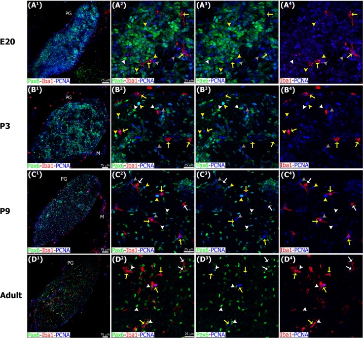
Fig 6. Microglia express the mitotic cell marker PCNA throughout the entire pineal gland ontogeny, while Pax6+ cells have detectable PCNA levels mainly during early development.
Panels show images of confocal microscopy of pineal gland (PG) sections immunostained for PCNA (blue), Pax6 (green) and Iba1 (red). The combination of these three markers revealed cell heterogeneity in the pineal gland in the different stages analyzed. Yellow arrows: Iba1/PCNA double-positive microglial cells. White arrows: microglia positive for Iba1 and negative for PCNA. Yellow arrowheads: Pax6/PCNA double-immunoreactive cells. White arrowheads: Pax6+ precursor cells negative for PCNA. Grey arrowheads: cells positive only for PCNA. (A1, C1, D1) 1.5x, 1.3x and 1.1x digital zooms from 10x images, respectively; (B1) 20x; scale bar: 75 μm. (A2-C4) 60x; (D2-D4) 1.4x digital zooms from a 40x image; scale bar: 25 μm. E, embryonic day. M, meninges. P, postnatal day.

