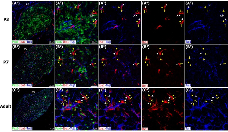
Fig 7. Microglia interact with Tuj1+ nerve fibers in the postnatal and adult pineal gland.
Sections of postnatal (P) and adult pineal glands (PG) immunolabeled for Pax6 (green), Iba1 (red) and β-tubulin III (Tuj1, blue). Sympathetic nerve fibers and microglia form an intricate network where Pax6+ cells are located. Macrophages (yellow arrows) phagocytosing Pax6+ cells (yellow arrowheads) and/or pruning nerve fibers (white arrowheads) are indicated. (A1-B1) 20x; (C1) 10x; scale bar: 75 μm. (A2-A5, B2-B5, C2-C5) 1.3x, 1.4x and 1.2x digital zooms from 60x images, respectively; scale bar: 25 μm.

