
Fig 11. Morphometric analysis shows that Pax6+ cell numbers decrease, while Iba1+ cell numbers, and Pax6+ cells engulfed by microglia increase in the pineal gland through ontogeny.
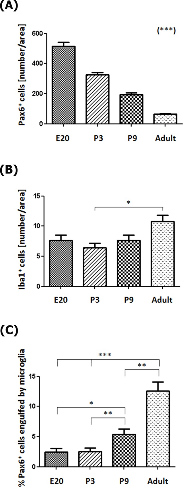
(A) The number of Pax6+ precursor cells per area (0.05 mm2) decreases throughout pineal development (Post-test for linear trend: P < 0.0001). (B) Microglia density increases slightly between postnatal day 3 (P3) and adulthood (Post-test for linear trend: P < 0.05). (C) The percentage of Pax6+ precursor cells in contact or engulfed by microglial cells is higher in late neonatal stages onwards (Post-test for linear trend: P < 0.0001). Data were expressed as mean ± S.E.M. Statistics: one-way ANOVA followed by the Tukey post-test: *** P < 0.001, ** P < 0.01, * P < 0.05. E, embryonic day.