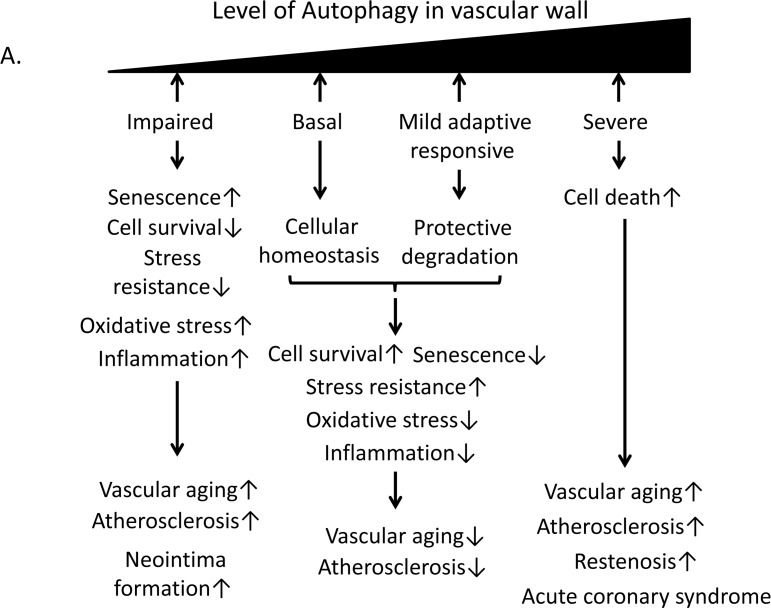
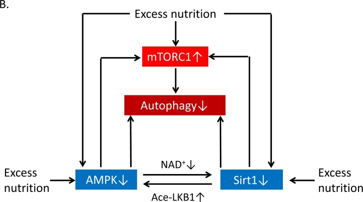
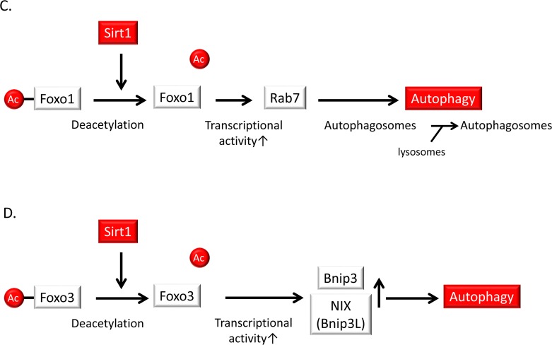
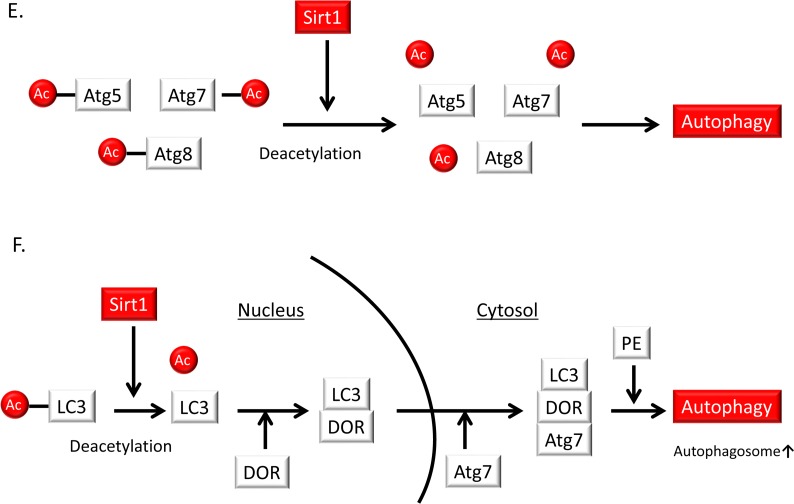
Figure 6. Relationship of autophagy levels to vascular aging and atherosclerosis and the role of Sirt1 in the regulation of autophagy.
(A) Basal autophagy and the mild adaptive autophagy response protect vascular cells against vascular aging and atherosclerosis through cellular homeostasis and protective degradation. By contrast, impaired or excessive autophagy leads to senescence, reduced stress resistance, oxidative stress, inflammation or cellular death, resulting in vascular aging and atherosclerosis. (B)Excess nutrition decreases autophagy initiation via mammalian target of rapamycin complex 1 (mTORC1) activation and reduced function of AMP-activated kinase (AMPK) and Sirt1. Nutrient-sensing pathways that include mTORC1, AMPK and Sirt1 engage in crosstalk with each other through changing of NAD+ level or acetylation/deacetylation of liver kinase B1 (LKB1). (C) Sirt1 induces the deacetylation, activation and nuclear translocation of FOXO1. FOXO1 activation stimulates the expression of Rab7, leading to the maturation of autophagosomes and autolysosomes via their fusion with lysosomes. (D) Sirt1 activation deacetylates and activates the FOXO3 transcription factor leading to subsequent Bcl-2/adenovirus E1B-19kDa interacting protein3 (Bnip3) or NIX/Bnip3-Like (Bnip3L)-mediated autophagy. (E) Sirt1 can form a molecular complex with several essential components of the autophagy machinery, including the autophagy-related genes (Atg) 5, Atg7 and Atg8. These autophagy components can be directly deacetylated by Sirt1. (F) Deacetylation of a nuclear pool of microtubule-associated protein light chain 3 (LC3) by Sirt1 initiates autophagy. Sirt1 deacetylates LC3 during starvation conditions. Deacetylated LC3 interacts with the nuclear protein DOR, and both proteins relocate from the nucleus to autophagosomes in the cytoplasm. Deacetylated LC3 interacts with Atg7 and other components of the ubiquitin-like conjugation machinery, leading to LC3 conjugation onto phosphatidylethanolamine (PE) and its incorporation into the early autophagosomal membrane.


