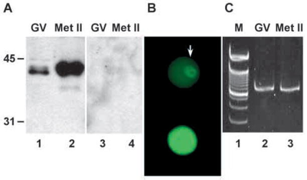
Fig. 1.
Expression of SLBP in oocytes and eggs. (A) Fifty germinal vesicle (GV)-stage oocytes and metaphase II (Met II) eggs were immunoblotted using anti-SLBP antiserum (lanes 1,2) or using anti- SLBP pre-incubated with the immunogenic peptide (lanes 3,4). Molecular weight markers are indicated to the left of the blots. (B) Oocytes were stained with purified anti-SLBP and analyzed by immunofluoresence. (Upper panel) GV-stage oocyte. The arrow indicates the nucleus. (Lower panel) Metaphase II egg. The photographs were taken using identical conditions. (C) Twenty-five GV-stage (lane 2) or metaphase II (lane 3) oocytes were subjected to RT-PCR using primers derived from the mouse SLBP cDNA sequence that were expected to amplify a fragment of 800 nt. Lane 1 shows 100-nt ladder; intense band near middle of gel is 700 nt. No signal was obtained when reverse transcriptase was omitted from the reaction (not shown).

