Figure 1.
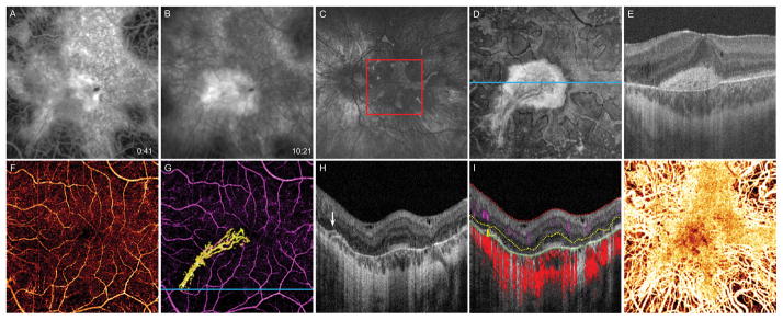
14-year-old boy with choroideremia complicated by CNV OS. A, 6 × 6 mm early-phase FA taken at diagnosis of CNV shows a central area of blockage, with, B, late-phase dye leakage. C, Fundus autofluorescence image 16 months after diagnosis shows diffuse chorioretinal atrophy with irregular perimacular sparing. Red box delineates 6 × 6 mm area corresponding to FA and OCT-A images. D, 16 months after diagnosis, en face OCT slab from 24 to 45 μm above BM, approximating the inner segment/outer segment junction, shows preserved photoreceptors at the macula. E, B scan corresponding to blue line in D illustrates that the central hyperreflective area in the en face image represents subretinal scar tissue with permeating vessels. F, OCT-A of the inner retinal vasculature shows preserved vessels. G, OCT-A of the inner retinal vasculature (purple) and CNV (yellow). H, B scan at the blue line in G reveals a bridging vessel (white arrow) from the choriocapillaris. I, Segmentation of the B scan illustrates flow at the location of the bridging vessel. Inner limiting membrane (red), outer boundary of the outer plexiform layer (yellow), and outer boundary of the RPE (green). Inner retinal circulation (purple shading), neovascular flow (yellow shading), and choroidal circulation (red shading). J, OCT-A of the choroid shows preserved choriocapillaris at the macula with deeper choroidal vessels visible at the periphery.

