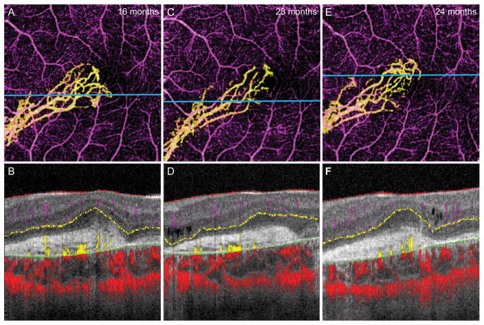
Figure 2.
14-year-old boy with choroideremia complicated by CNV OS. A, 3 × 3 mm OCT-A of the isolated CNV 16 months after diagnosis, with B, the segmented B scan at the level of the blue line shown in A. C, 3 × 3 mm OCT-A of the CNV 23 months after diagnosis shows diminished capillary branching in the inferior temporal region but increased growth at the temporal tip. D, Segmented B scan at the level of the blue line shown in C. E, 3 × 3 mm OCT-A of CNV 24 months after diagnosis, with F, the segmented B scan at the level of the blue line shown in E, which appears similar to the previous scan.

