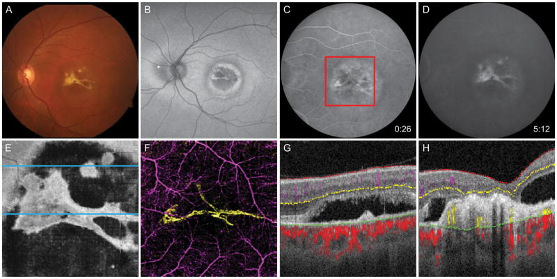
Figure 4.
60-year-old man with Best vitelliform dystrophy complicated by CNV OS. A, Fundus photo shows yellow region at macula with bright lesion resembling fibrosis. B, Fundus autofluorescence demonstrates alternating concentric rings of hyperautofluorescence and hypoautofluorescence at the macula. C, Early-phase FA reveals mottled early hyperfluorescence, and D, late staining. E, En face OCT slab at the inner segment/outer segment junction demonstrates subretinal fluid containing subretinal fibrosis. F, 6 × 6 mm OCT-A corresponding to the red box in C demonstrates the CNV (yellow) is composed of two broad loops with minimal capillary networks. G, Segmented B scan at the superior blue line in E reveals avascular fibrotic tissue and subretinal fluid but no detectable flow. H, Segmented B scan at the inferior blue line in E shows angiographic flow within fibrotic tissue.

