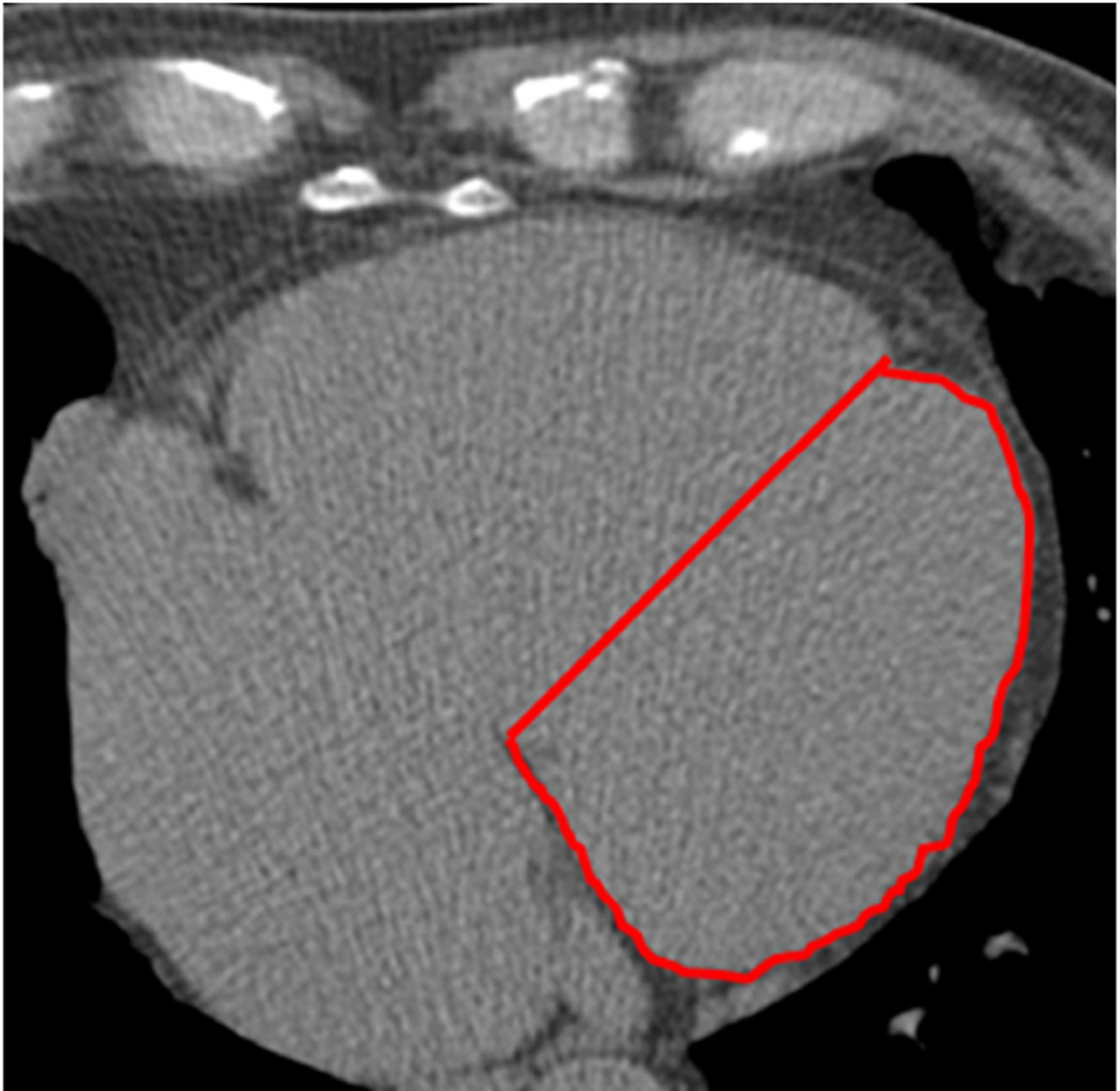
Figure 1. Example of the LVA measurement.
The antero-posterior juncture origin is the interventricular groove, identified by the natural markers, such as the abrupt dip that represents fat tissue. The lateral border is easily identified by the abrupt change in contrast density from the cardiac silhouette to the pericardial fat. The posterior reference is the atrioventricular groove. A straight line connecting the antero-posterior juncture of both ventricles is drawn to complete the tracing (red line).

