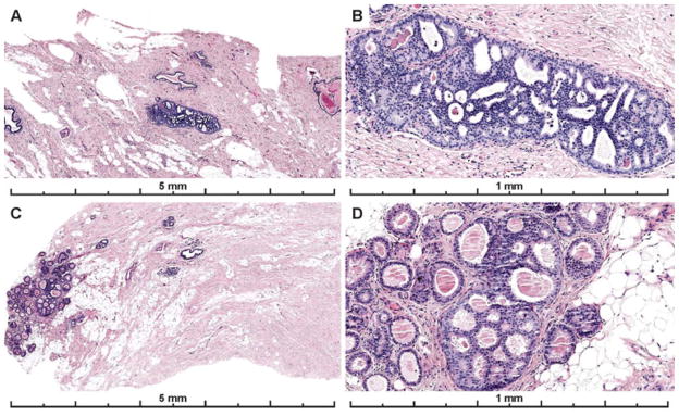
Figure 3.
Photos of the diagnostic areas from the two cases with the highest participant agreement with the consensus diagnosis of ADH. Both cases had focal lesions, less than 2 mm (and < 2 membrane bound spaces), that were obvious on low power, had obvious cytologic monotony and a cribriform architectural pattern. A–B) A case with 83% of participant agreement with the expert consensus diagnosis of ADH; 7% recorded a Benign diagnosis (UDH), and 10% recorded a DCIS diagnosis for the case (30 total interpretations). C–D) 89% agreed with the consensus diagnosis of ADH; 4% recorded a Benign diagnosis (UDH) and 7% recorded a DCIS diagnosis on the case (27 total interpretations). Lesions with these features should be reproducibly classified as ADH as serve as good examples of this diagnosis.

