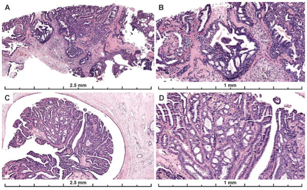
Figure 4.
Papillary lesions with ADH were associated with higher diagnostic agreement than ADH not in a papillary lesion. A–B) 67% of participants recorded a diagnosis of ADH for this papillary lesion; 28% recorded a DCIS diagnosis and 3% a Benign diagnosis (mostly papilloma without atypia) (29 interpretations). C–D) 59% of participants recorded a diagnosis of ADH for this papillary lesion; 31% recorded a Benign diagnosis (mostly papilloma without atypia), and 10% recorded a DCIS diagnosis for the case (29 total interpretations). The first case (A–B) had less discrete areas of atypia in more than one area of the papilloma, making it borderline with DCIS. However, each focus was considered < 2mm and the consensus diagnosis was ADH on this core needle biopsy sample. The second case has a single, more discrete area of cribriform architecture with cytologic monotony within the papillary proliferation measuring < 2 mm. These areas appear distinct from the background normal papillary epithelium or UDH but due to their limited in extent (< 2 mm), they fall short of most pathologists’ threshold for DCIS.

