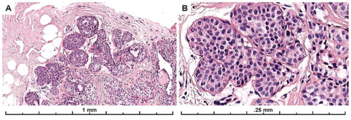
Figure 6.
Example images of the diagnostic area from a case with solid architecture and subtle cytologic monotony. Only 17% of participating pathologists recorded a diagnosis of ADH, with 55% recording the case as LN and 28% as DCIS (29 total interpretations). While the differential diagnosis includes a lobular in situ lesion (ALH/LCIS) that may be resolved with an E-cadherin stain, subtle micro-acini supporting a ductal process are evident on the H&E at high power (panel B).

