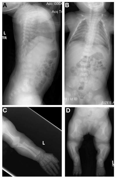
FIG. 1.
Radiographs of the proband at age 22 months. (A and B) Lateral and anterior–posterior images of the spine. Platyspondyly with anterior rounding can be seen in both images with moderate scoliosis apparent in (B). (C) Upper extremity showing shortened long bones with wide metaphyses and short phalanges. (D) Lower extremities showing short long bones with widened metaphyses, halberd-shaped proximal femurs as well as the abnormal pelvis characteristic of metatropic dysplasia.

