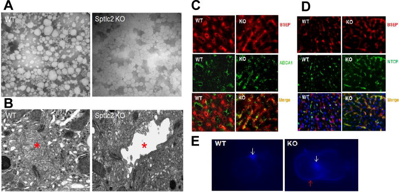
Figure 3. Impairment of Hepatocyte Apical-basal Polarity.
(A) Electronic microscope images of mouse plasma lipoproteins which were negative stained. (B) Electronic microscope images of mouse liver sections. Bile canaliculus was indicated by red asterisks. (C) Bile salt export pump (BSEP) and ABCA1 were immuno-stained on liver sections. (D) BSEP and sodium-taurocholate co-transporting polypeptide (NTCP) were immuno-stained on liver sections. (E) Freshly isolated hepatocyte couplets were stained with Filipin. White arrows indicate bile canaliculus and red arrow indicates basal membrane. The pictures are the representatives of three mice/group.

