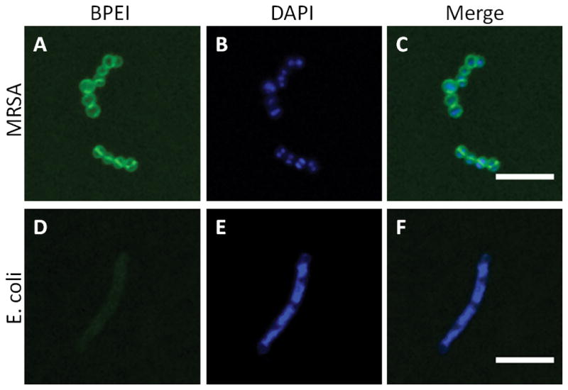
Figure 2.

Optical sections of BPEI binding to MRSA and E. coli. Paraformaldehyde-fixed MRSA, stained with BPEI-Alexa Fluor 488 (A) and DAPI (B), is imaged by LSCM. The merged image (C) shows BPEI binding to the cell surface but not within the cytoplasm. In contrast, PFA-fixed E. coli stained with BPEI-Alexa Fluor 488 (D) and DAPI (E), and the merged image (F), shows a relatively low affinity between BPEI and E. coli. Scale bar = 5 μm.
