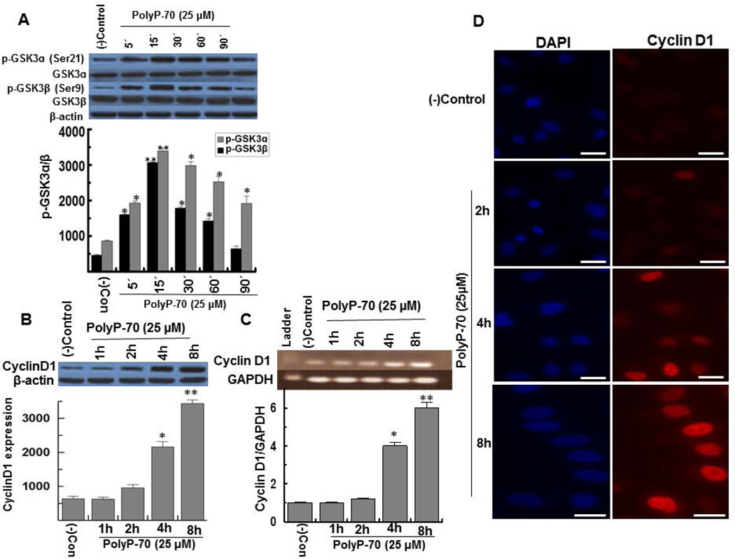
Figure 1.
PolyP-70 induces GSK-3 phosphorylation and increases expression and nuclear localization of cyclin D1, in EA.hy926 endothelial cells. (A) Time course of polyP-mediated phosphorylation and inactivation of GSK-3α/β in EA.hy926 cells. (B) Cells were treated with polyP-70 (25 µM) at different time points followed by measuring the expression of cyclin D1. (C) Semiquantitative RT-PCR was performed as described before (22). Results were normalized to expression levels of GAPDH and presented as the fold difference relative to the control group at each time point. The results are shown as mean ± standard deviation of 3 different experiments as determined Student t test. *P<0.05; **P<0.01. (D) The same as B except that the effect of polyP-70 on nuclear localization of cyclin D1 was measured. Cells were stained with DAPI to visualize the nucleus (Blue) and anti-cyclin D1 antibody (Red) and then imaged by confocal microscopy. The scale for the microscopic figure is 20µm.

