Figure 1.

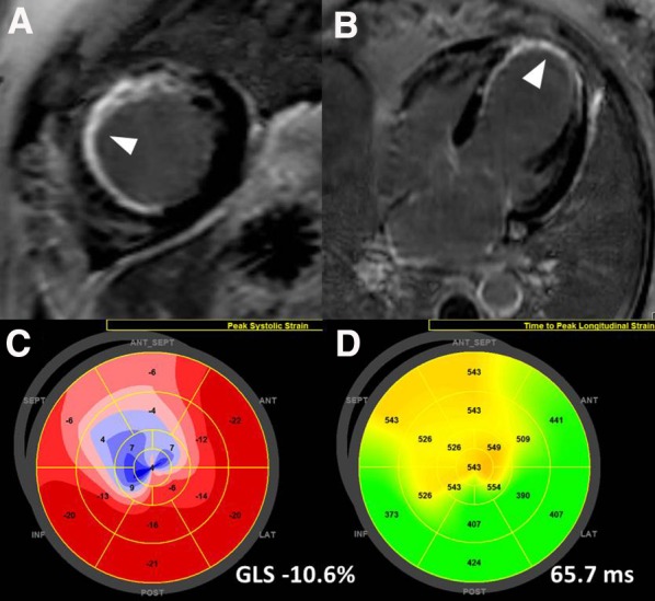
Cardiac magnetic resonance and echocardiographic speckle tracking analysis for risk stratification of patients with ischemic heart disease. Panels A and B show transmural myocardial scar in the apical septal and anteroseptal segments (arrows) and subendocardial scar in the mid-inferoseptal segment. On 2-dimensional speckle tracking echocardiography, the magnitude of global longitudinal strain is −10.6% (panel C). The LV apical segments show positive values and are color coded in blue indicating lengthening (correlating with the area of transmural scar). Panel D shows significant mechanical dispersion (65.7 ms) based on the standard deviation of time to peak longitudinal strain of 17 segments. The most delayed areas coincide with the areas with scar and impaired longitudinal strain
