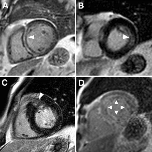
Figure 2.

Patterns of late gadolinium contrast enhancement in nonischemic cardiomyopathies. Septal mid-wall late gadolinium enhancement (arrow) is typically observed in dilated cardiomyopathy (A). Mid-wall late gadolinium enhancement of the basal inferolateral wall (arrow) in a patient with cardiac sarcoidosis (B). Patchy mid-wall late gadolinium enhancement of the hypertrophic septum at the level of the right ventricular junction (arrow) is typical of hypertrophic cardiomyopathy (C). In cardiac amyloidosis (D), the pattern of late gadolinium enhancement is characterized by circumferential subendocardial distribution (arrows)
