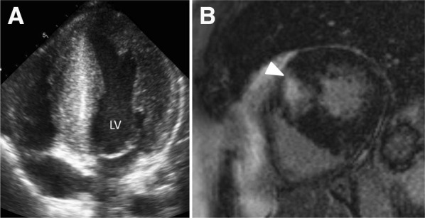
Figure 3.

Hypertrophic cardiomyopathy. Panel A shows left ventricular (LV) hypertrophy with >15 mm thickness of the septal and lateral walls. Panel B shows late gadolinium-enhanced cardiac magnetic resonance of a patient with hypertrophic cardiomyopathy and delayed enhancement in the septum, at the insertion of the right ventricle (arrow)
