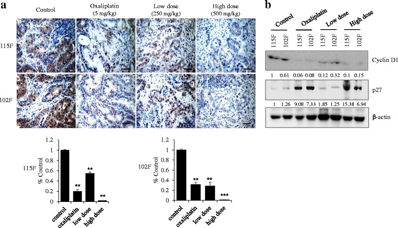
Fig. 4.

Inhibition of cell proliferation by BP3B in PDTX model. a To check the effect of BP3B on cell proliferation, immunohistochemical analysis of Ki67 was performed with drug-treated samples. Ki67 were stained into brown and nuclei were counterstained with hematoxylin (purple). The intensity of Ki67-positive cell was calculated with ImmunoRatio software. Scale bars are 50 μm. * p < 0.05, ** p < 0.01, *** p < 0.001. b Expression of cell proliferation markers (Cyclin D1 and p27) was confirmed with western blot. Densitometric analysis was performed with Image J software
