
Fig. 4.
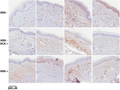
C3b staining of skin biopsied from anti-RNA polymerase III (ARA+), ARA− and anti-centromere-positive (ACA+)/ARA− patients. Shown are representative micrographs of skin biopsies immunostained for C3b. Four patients per group (rows) were available for the study. In most images, the endothelium of the dermal arterioles is distinctly positive. There is also a fainter staining associated with the dermal collagen strands. In the basal layer of the epidermis, melanocytes are seen, filled with melanin pigment but negative for C3b