
Figure 1.
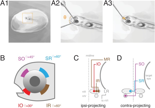
Labeling of extraocular motoneurons by retro‐orbital dye fill. (A) Retro‐orbital fill procedure. Larvae were anesthetized and immobilized in 2% agar, target eye near the surface (A1). Agar was cleared from eye and a tungsten needle used to make an incision of angle (Θ) in the vicinity of one/more extraocular muscle (A2). Crystallized dye was placed at incision site (A3); somata were imaged later. (B) Targeted position and angle (Θ) of incision around eye to label a given motoneuron population. (C) Schematic of extraocular muscle innervation by ipsilaterally projecting motoneurons. LR motoneurons located in nVI were not targeted for dye fills. (D) Schematic of extraocular muscle innervation by contralaterally projecting motoneurons. MR, IR, SR, LR: medial, inferior, superior, lateral rectus. SO, IO: superior, inferior oblique. nIII: oculomotor nucleus. nIV: trochlear nucleus. nVI: abducens nucleus.