
Figure 5.
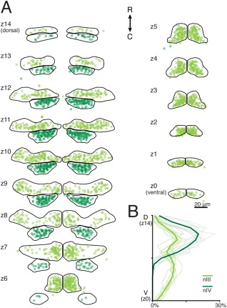
Distribution of labeled motoneuron somata in 5–7‐day‐old Tg(isl1:GFP) zebrafish. (A) GFP+ motoneuron somata are shown as circles in nIII (light green) and nIV (dark green). The dorsoventral extent of nIII/nIV is subdivided into 15 6‐μm‐thick planes, labeled z14 (most dorsal) – z0 (most ventral). (B) Mean and individual (background traces) probability distributions from labeled motoneurons across nIII/nIV. nIII: n = 10 larvae, 2080 cells. nIV: n = 10 larvae, 936 cells.