Fig. 2.

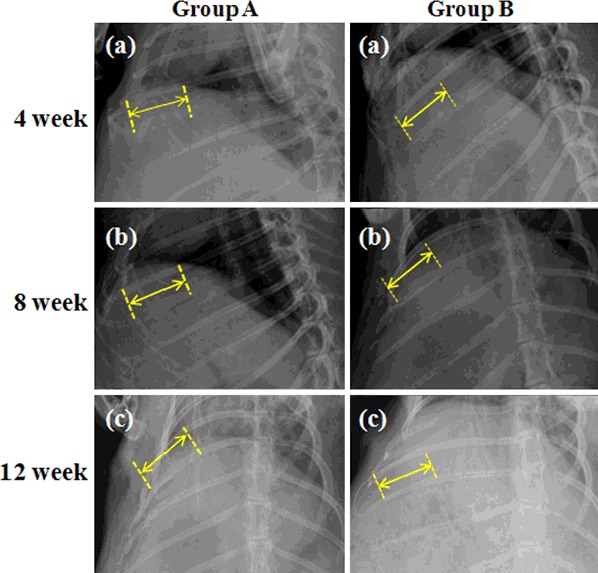
X-ray radiographs of rabbit rib defects at 12 weeks after surgery. Group A: blank control group, MC was not implanted in the rib defect. Group B: MC group, MC was implanted in the bone segment (3 cm defect)

X-ray radiographs of rabbit rib defects at 12 weeks after surgery. Group A: blank control group, MC was not implanted in the rib defect. Group B: MC group, MC was implanted in the bone segment (3 cm defect)