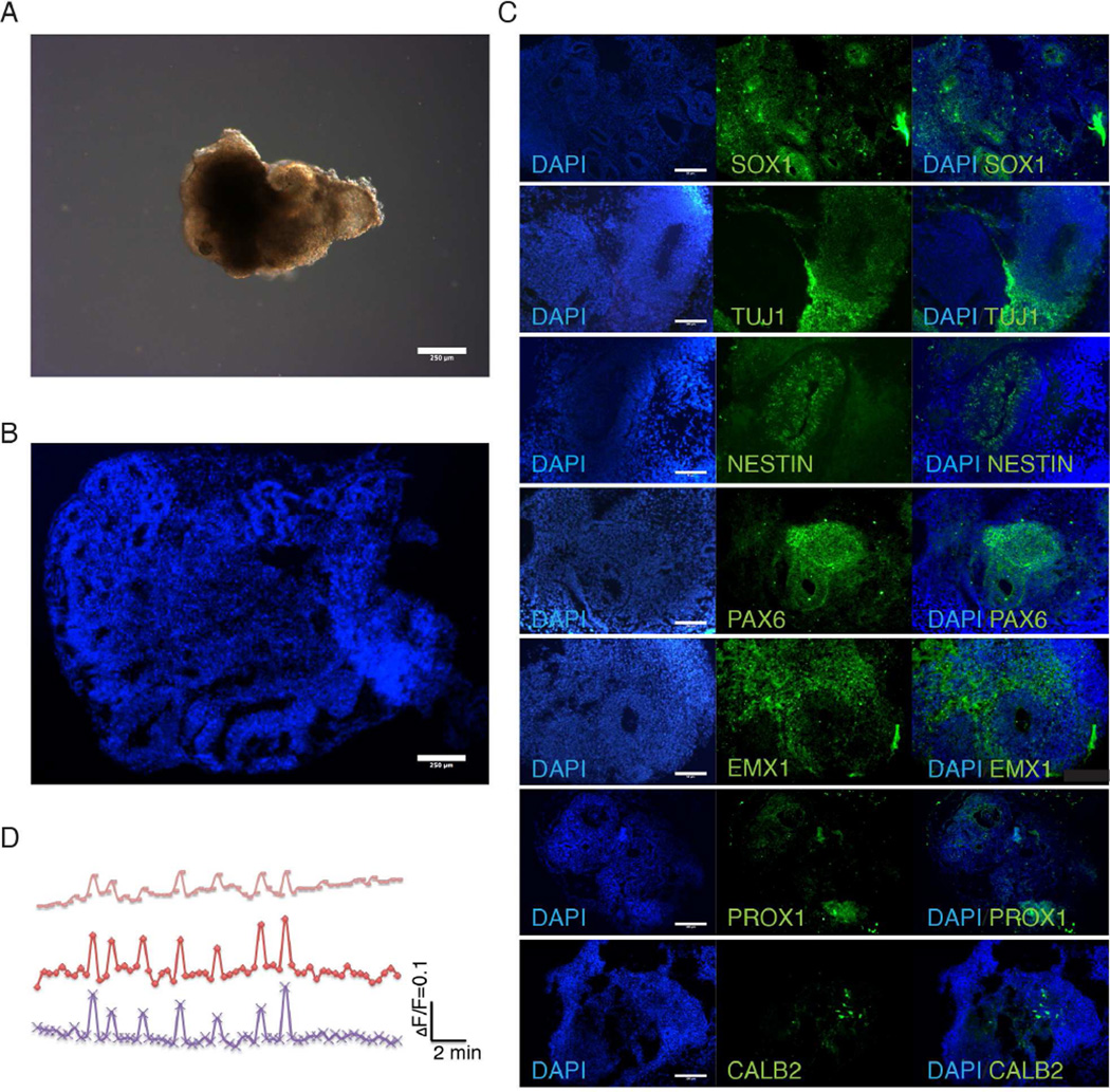
Figure 1.
Characterization of cerebral organoids reveals recapitulation of fetal brain regions. (A) Bright-field image of representative organoids show development of neuroepithelial layer. Scale bar: 250µm. (B) DAPI stained organoid shows complex inner morphology including ventricle-like structures from 30-day-old organoids. Scale bar: 250µm. (C) Organoids immunostained for neuronal (TUJ1+) and neural progenitor cells (SOX1+) cells. TUJ1 shows generalized neuronal differentiation while neural progenitors are localized near inner ventricle-like structures in 30-day-old organoids. Immunostaining for forebrain (PAX6), dorsal cortex (EMX1), hippocampus (PROX1) and interneurons (CALB2) show differentiation of organoids into discrete brain regions 30-day-old organoids. Also see Figure S1. Scale bar: 100µm. (D) Calcium dye imaging of cerebral organoids using Fluo-4 shows functional neural activity.

