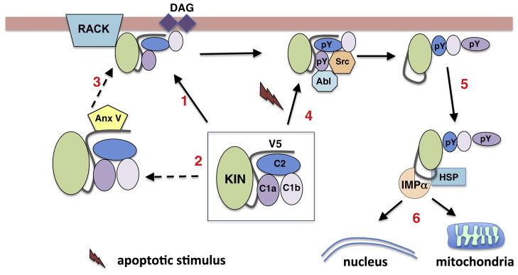
Fig. 2.
Mechanisms of PKCδ activation. PKCδ is maintained in an inactive state through a series of interactions between the kinase and regulatory domains (C1, C2a, and C1b). Activation can occur through multiple pathways. In response to generation of DAG PKCδ translocates to cellular membranes (arrow1). This may occur through interaction with scaffolding proteins such as RACKs (also AKAPs, C-KIPS, and p23/Tmp21; see text for more information). In some cases, translocation requires interactions with other cellular factors such as Annexin V (arrows 2 and 3). Non-canonical activation of PKCδ can result from upstream activation of Abl resulting in increased phosphorylation of Y155 (pY) in the C1a domain, which results in phosphorylation of Y64 (C2 domain) by Src kinase (arrow 4). This exposes a nuclear localization sequence that binds importin-α leading to nuclear import (arrows 5 and 6). Alternatively, PKC can traffic to mitochondria through interactions with HSP family members and Abl (arrow 6).

