Fig. 1.


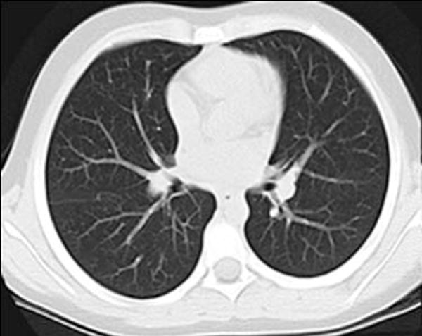
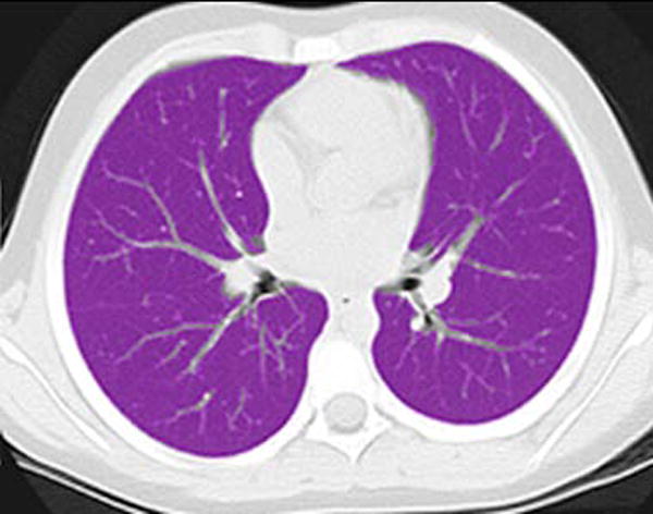
Lung segmentation technique. a Standard gray-scale non-contrast axial CT image through the mid-thorax in a 69-month-old girl. b Corresponding magenta-shaded mask highlights the segmented lung parenchyma


Lung segmentation technique. a Standard gray-scale non-contrast axial CT image through the mid-thorax in a 69-month-old girl. b Corresponding magenta-shaded mask highlights the segmented lung parenchyma